RedefiningProstate Imaging.
Revolutionizing prostate MRI diagnosis
with Artificial Intelligence.
Funded by

Revolutionizing prostate MRI diagnosis
with Artificial Intelligence.
Funded by
Combined Efforts with


Advanced Prostate Analysis
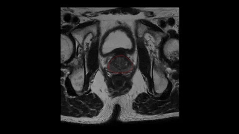
AI-assisted Prostate Cancer Diagnosis
Comprehensive diagnosis powered by state-of-the-art vision transformers
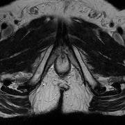
Manual Clinical Adjustments
Radiologist-guided adjustments with real-time 3D visualization
Intuitive mesh editing tools to fine-tune AI segmentations
Real-time 3D preview for precise anatomical corrections
Empowers expert oversight for clinical-grade accuracy
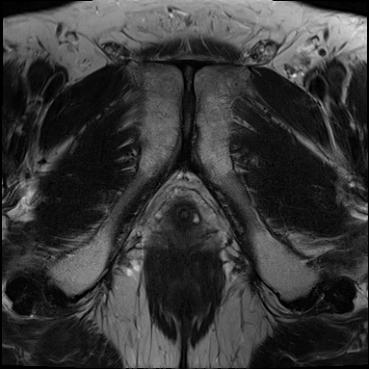
segmentation
slice: 01/18segmedix-unet
segmedix-conv
segmedix-vit
⚡
SOTA Prostate Segmentation Models
State-of-the-art AI architectures for optimal segmentation performance
segMedix-UNet: Fast and accurate for standard T2W images
segMedix-Conv: Enhanced edge detection for complex cases
segMedix-ViT: State-of-the-art transformer-based analysis
Lesion Detection
Automated identification and classification of suspicious lesions
AI-powered lesion localization and analysis
PI-RADS scoring calculator for clinical workflow
Lesion segmentation mapped to prostate zones and regions
Anatomically-aligned output for fusion biopsy integration
Automated Documentation
Clinical Reporting
Generate comprehensive clinical reports with quantitative measurements and structured findings
Formats
MultiExport
EHR/RIS
FullyCompatible
Clinical Report
PDF
XML
DICOM SR
Report Ready
Multi-format Export
Seamless Integration
PACS Integration
Direct integration with your existing PACS system. No workflow disruptions, no data silos, just seamless image access and collaboration.
HIPAA
CompliantPrivacy & Security
DICOM
SupportedImaging Standard
PACS Server

segMedix AI
Setup <2 hours
ExperiencesegMedix.
Experience AI-powered prostate imaging
with a personalized demonstration.
Questions?
Contact our team →